FOR IMMEDIATE RELEASE
ACS News Service Weekly PressPac: June 03, 2015
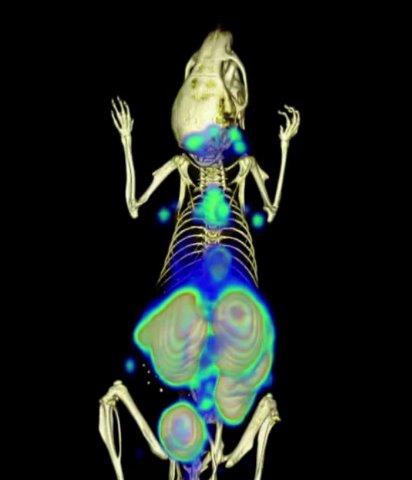
Antibody fragments expand what PET imaging can ‘see’ in mice (video)
"Use of 18F‐2-Fluorodeoxyglucose To Label Antibody Fragments for Immuno-Positron Emission Tomography of Pancreatic Cancer"
ACS Central Science
To visualize cancer throughout the body, physicians often turn to positron emission tomography (PET), which lights up areas that are metabolically active or growing, like tumors. Today in ACS Central Science, researchers report development of new PET probes composed of labeled antibody fragments that were tested in mice. These probes could someday be used to create targeted probes, giving doctors more information about tumors and how to treat them.
The most common PET imaging probe is a labeled sugar molecule called 18F-2-deoxyfluoroglucose (FDG). PET indicates those locations where a labeled FDG molecule ends up. Because tumor cells grow faster than healthy ones, they need more sugar to meet their energy requirements, and they consume much more of the labeled sugar probe. This makes PET ideal for seeing tumors. But this method is limited because the reporter part of the molecule, 18F, is difficult to attach to other molecules and isn’t very stable. FDG molecules are also general probes, since they can be taken up wherever the body needs energy, leading to background signals. Hidde Ploegh and coworkers set out to overcome these challenges by making probes that meet all of these needs.
The researchers developed a method to outfit antibody fragments with FDG. To demonstrate the usefulness of this approach, they linked a labeled FDG molecule to an antibody fragment to detect pancreatic cancer. The resulting immuno-PET probe was able to visualize pancreatic cancer tumors too small to be detected in mice using the labeled FDG molecule alone. Ploegh and coworkers conjecture that the new probes could be used in concert with the conventional ones to test both the biochemical and metabolic signatures of cancer in patients. This information could help doctors decide between different treatment options, or even to visualize the delivery of drugs.
The authors acknowledge funding from the National Institutes of Health and the Lustgarten Foundation.
A video of the PET probe in action is available at https://youtu.be/xJTnaAy5y5c.


